
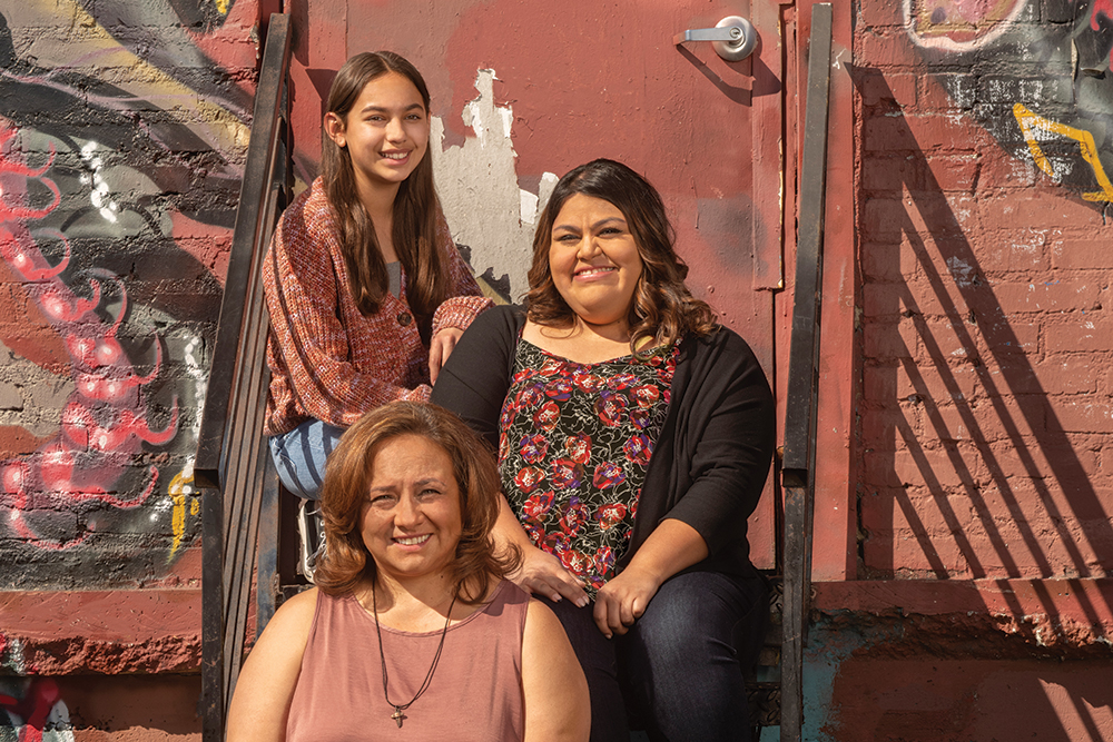
The technology found a precancerous lesion in Christine Ysabal. After a surgery to remove it, she has become an advocate for regular screenings to inspire her daughters.
As a kid, Christine Ysabal, JD, LLM, thought “doctors’ appointments were only for when you were ill,” she says, not for preventing future health problems. She had routine vaccinations but never saw anyone in her family get an annual checkup or screening.
So, when she turned 40 and people told Christine she should start having a yearly mammogram, she put it off.
A busy mom and wife, she was studying for her master’s degree and working full-time as a contracts and compliance administrator in the Department of Radiology at Keck Medicine of USC.
“Everything got prioritized ahead of my own health,” she says.
Christine finally made time for her first mammogram at age 46, and everything was fine. With that good report and no family history of breast cancer, Christine didn’t schedule another screening until four years later in October 2020.
Why all women should get a 3D mammogram
At that time, USC Norris Comprehensive Cancer Center diagnostic radiologist Mary Yamashita, MD, ordered a 3D mammogram for Christine due to her dense breast tissue. In any mammogram, dense breast tissue appears white on the image.
The problem? “Breast cancer also appears white, making it more difficult to find small cancer in women with dense breast tissue,” Dr. Yamashita says.
The 3D mammogram technology produces multiple X-ray images from multiple angles, then reconstructs them into one-millimeter slices. Evaluating the breast 1 mm slice at a time can allow clinicians to detect cancers hidden by overlying dense breast tissue.
Keck Medicine has offered 3D mammograms — also known as digital breast tomosynthesis — since its approval by the U.S. Food and Drug Administration in 2011.
“We’re able to identify more cancer and smaller cancers as a result,” says Dr. Yamashita, a clinical associate professor of radiology at the Keck School of Medicine of USC.
She recommends that every woman — whether at average risk or high risk for breast cancer, with dense breast tissue or fatty tissue — get a 3D mammogram.
3D mammogram detects hard-to-see breast tumor
For Christine, the technology was a lifesaver. In the detailed images, Dr. Yamashita saw something that concerned her: a suspicious-looking lesion.
“I don’t know how I missed the lump,” Christine says.
A follow-up diagnostic ultrasound focused on the area of concern seen on the mammogram revealed an abnormal mass, as Dr. Yamashita suspected. A needle biopsy then confirmed that Christine had a “precancerous” tumor that could develop into cancer.
“Breast screening enables us to find cancers before you can feel it,” Dr. Yamashita says.
Since 1989, when mammograms became widely available in the U.S., through 2015, the breast cancer death rate has dropped 43%.
“We want every woman to get screened because breast cancer when detected early is curable,” she adds. “And you require less treatments, less surgery, and less toxic chemotherapy.”
In 2020 and early 2021, under the cloud of COVID-19, some women postponed their mammograms. “In a few cases, we found cancer and wondered if we would have caught it smaller if the women had come in the year before,” Dr. Yamashita says.
Regular breast screenings catch problems early
In November 2020, Christine had a lumpectomy, a surgical procedure to remove the lump from her breast. Because the lesion was not cancerous, Christine didn’t need additional treatment.
For the foreseeable future, she will have yearly checkups with a breast surgeon and two imaging studies annually — a mammogram and magnetic resonance imaging (MRI). These regular exams will ensure any future tumors will be caught early, such as the benign mass that was removed in April 2022.
Put yourself first. Get screened.”
Christine Ysabal, patient, USC Norris Comprehensive Cancer Center
Which type of imaging is recommended — mammogram, ultrasound, and/or MRI — depends on the individual patient’s risk factors.
“This goes beyond personalized medicine,” Dr. Yamashita says. “This is precision medicine.”
Recently, Dr. Yamashita led Keck Medicine’s participation in a multi-center clinical trial studying fully automated breast ultrasound for women with dense breast tissue, which won FDA approval to broaden cancer surveillance options.
Christine advises others: “Put yourself first. Get screened.” She is determined to be a positive role model for her daughters: Isabella, 12, and Samantha, 32.
“I want to be around and healthy in their lives for a long time,” Christine says.
Topics